What Is Hyperspectral Imaging?
Hyperspectral imaging is a discipline that uses specialized sensors to capture data at numerous narrow wavelengths simultaneously. Hyperspectral data is typically represented as an image cube in which each image represents one of the tens or hundreds of narrow wavelength ranges or spectral bands. Hyperspectral imaging enables measurement and analysis of the spectral characteristics of regions or objects, which are used in applications like agricultural crop health assessment, environmental degradation monitoring, and tissue pathology.
Where Is Hyperspectral Imaging Used?
Hyperspectral imaging is used in applications like remote sensing, surveillance, machine vision, and medical imaging with the primary purpose of identifying materials and detecting objects, anomalies, and processes. For example, hyperspectral imaging in remote sensing involves detecting and identifying minerals, terrestrial vegetation, and man-made structures using satellite or aerial images.

Using satellite-based hyperspectral data to identify different terrestrial regions, based on their material type, using maximum abundance classification.
In medical imaging, hyperspectral imaging analysis of tissue surfaces can provide insightful diagnostic information for tissue pathology.

Hyperspectral images from The University of Nottingham, derived by shining light on the tissue surface and accurately measuring oxygen levels and generating oxygen saturation maps to facilitate the work of clinical researchers and doctors.
How Does Hyperspectral Image Processing Work?
Hyperspectral imaging begins with capturing a set of images using hyperspectral image sensors and representing them as a three-dimensional hyperspectral data cube. This data cube can then be analyzed and processed using a variety of image processing algorithms, mainly for workflows involving classification, spectral matching, and unmixing.

A typical hyperspectral image processing workflow, which involves representing, analyzing, and interpreting information contained in the hyperspectral images.
For example, a common hyperspectral image processing technique, maximum abundance classification (MAC), is used to classify pixels in an image based on their spectral signatures, and associate them to an object or region class, also known as endmember class.

An abundance map generated using maximum abundance classification of a hyperspectral image with nine endmember classes: Asphalt, Meadows, Gravel, Trees, Painted metal sheets, Bare soil, Bitumen, Self-blocking bricks, and Shadows.
Another common task involves using the Normalized Difference Vegetation Index or NDVI values of hyperspectral image of a vegetation region and applying a threshold on this value to measure the relative health of vegetation across this region. The higher the NDVI value, the healthier and denser the vegetation is in that region.

The (right-most) image with highest NDVI value has indicated regions with healthier and denser vegetation in green.
Hyperspectral Imaging with MATLAB
The Hyperspectral Imaging Library in MATLAB® provides a set of functions and visualization capabilities for accessing, preprocessing, analyzing, and processing hyperspectral data.
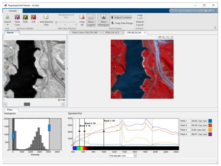
The Hyperspectral Viewer app enables visualization and interactive exploration of the hyperspectral data. You can view the individual bands of the hyperspectral data as gray-scale images as well as the color composite representations of the data. The app also enables creating spectral profiles that enable identifying the constituting elements in the hyperspectral data.

Hyperspectral Viewer app for visualizing hyperspectral data and spectral profiles.
The Hyperspectral Imaging Library contains built-in functions for preprocessing, analyzing and interpreting hyperspectral data. It supports workflows for dimensionality reduction, spectral unmixing, spectral matching, and classification with techniques such as band selection, endmember extraction, abundance map estimation, NDVI, and anomaly detection.

Endmember extraction and abundance map estimation for spectral unmixing.
MATLAB provides tools for data exploration, visualization, and algorithm development that support hyperspectral imaging workflows. In addition, Image Processing Toolbox™ provides a comprehensive set of reference-standard algorithms that you can use with hyperspectral imaging data.

MATLAB used for plotting and visualizing the spectral match score maps.
